- Institution: Stanford Univ Med Ctr Lane Med Lib/Periodical Dept/Rm L109
- Sign In as Member / Individual
PROMISCUOUS LIGANDS AND ATTRACTIVE CAVITIES

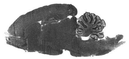
Figure 8.
Autoradiogram of rat brain section photoaffinity labeled with radioactive halothane. Degree of halothane binding is indicated by level of darkness; no other staining has been applied to the section. The binding shows little regional preference and is reduced to near-background levels in the presence of a tenfold excess of unlabeled halothane. The dramatic inhibition of labeling by non-radioactive halothane indicates that most halothane binding is saturable and specific, showing that many proteins could be involved in anesthetic action [from (45)].


