- Institution: Stanford Univ Med Ctr Lane Med Lib/Periodical Dept/Rm L109
- Sign In as Member / Individual
The “Heartbreak” of Older Age

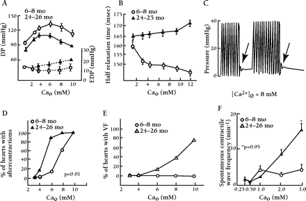
Reduced Ca2+tolerance of senescent hearts. A. Developed pressure (DP) and end-diastolic pressure (EDP) in response to increasing perfusate Ca2+ concentration {[Cao ]} in young and old hearts during stimulation at 2.0 Hz. Responses of both DP and EDP significantly differ with age by repeated-measures analysis of variance. B. Half-relaxation time in young and old hearts in response to an increase in [Cao ] at 2.0-Hz stimulation. Response differs with age. C. Chart recording of left ventricular pressure during and after cessation of electrical stimulation. Note the occurrence after cessation of stimulation in 8 mM [Cao ] of a “hyperrelaxation” and aftercontractions (arrows), followed by a slow decay in diastolic pressure, which eventually reaches the RP level (not shown) D. Aftercontractions occurrence after cessation of electrical stimulation at 2.0 Hz in young and old hearts as a function of [Cao ]. E. Spontaneous ventricular fibrillation (VF) occurrence during pacing at 2.0 Hz as a function of [Cao ]. F. Spontaneous contractile waves generated by spontaneous Ca2+ oscillations in single isolate ventricular myocytes from senescent hearts occur more frequently than in those from young adult hearts as [Cao ] is increased. From (16).


