
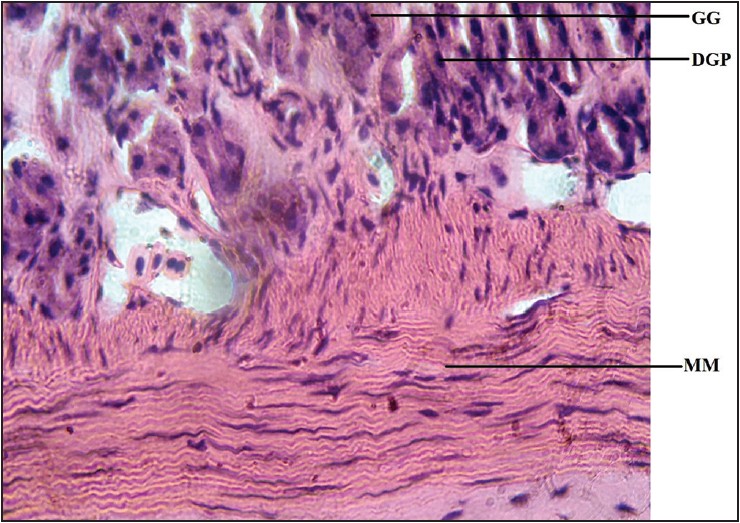
Figure 8: Photomicrograph of cane toad stomach showing gastric gland (GG), muscularis mucosae (MM), dark pigmented granule (DPG). Stained with H&E (400×)
| Close | |
|
|
|
|
Figure 8: Photomicrograph of cane toad stomach showing gastric gland (GG), muscularis mucosae (MM), dark pigmented granule (DPG). Stained with H&E (400×)
|
|