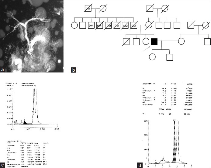
Figure 1: (a) Contrast enhanced computed tomography image of proband showing necrosis in pancreas, (b) Pedigree of family showing family history of diabetes mellitius type II, (c) HbA1c chromatogram of proband showing abnormal pattern, (d) HPLC chromatogram of proband showing presence of HbE and HbD