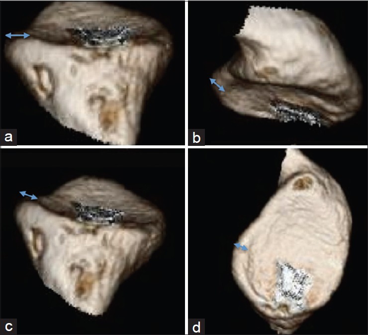
Figure 2: Various views from 3D CT reconstruction of an uninjured glenoid showing the presence of bone beyond the peak of the anterior glenoid rim. (a) View from inferior to superior; (b and c) Views from superior to inferior; (d) en-face view of glenoid
