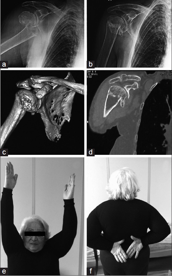
Figure 2: A 79-year-old female patient with Neer type two-part fracture. (a) Initial shoulder antero-posterior shoulder radiograph. (b) Final follow-up radiograph. (c) 3D computerized tomography (CT) and (d) coronal CT reconstruction. (e and f) Clinical appearance of the patient at the final follow-up at 22 months
