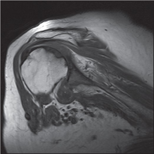
Figure 2: T2 weighted coronal MRI scan showing abnormal increased signal in the distal supraspinatus tendon which appears to be thinned and compressed with no definite tear or tendon retraction seen


|
Close |
|
Figure 2: T2 weighted coronal MRI scan showing abnormal increased signal in the distal supraspinatus tendon which appears to be thinned and compressed with no definite tear or tendon retraction seen
|
|