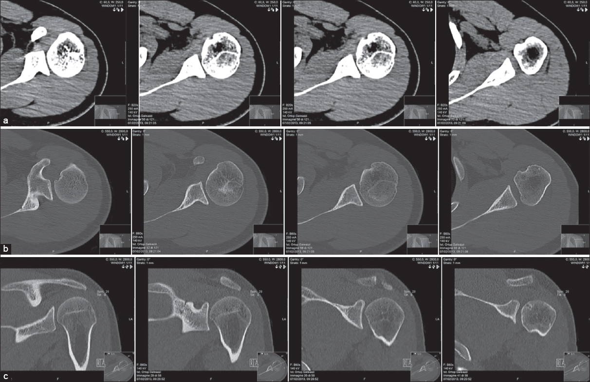
Figure 2: Pre-operative computed tomography images. Axial and sagittal view. Left shoulder: humeral head that presents focal cortical erosion with small subcortical cystic-dystrophic irregularities along the anterior superior medial side. Incision of the profi le cortex of the humeral head on the rear side. (a-b) Axial view (c) sagittal view