
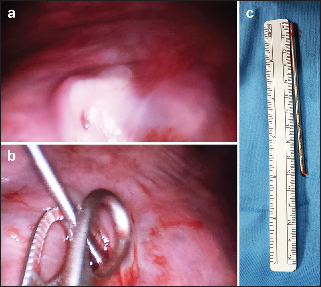
Figure 3: (a) Fibrin-coated entry point of the wire in the left lung; (b) Thoracoscopic views of the wire removal; (c) The removed wire
|
|
Close |
|
Figure 3: (a) Fibrin-coated entry point of the wire in the left lung; (b) Thoracoscopic views of the wire removal; (c) The removed wire
|
|