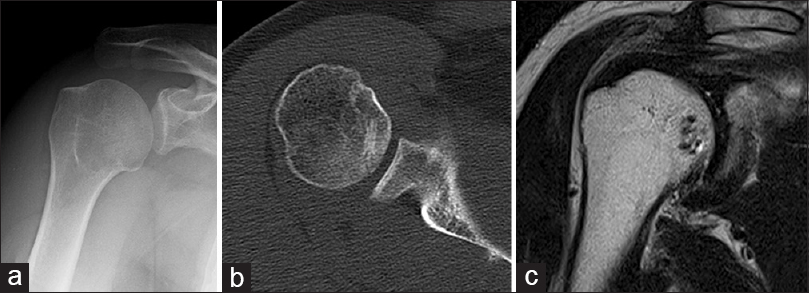
Figure 3: The imaging and arthroscopic findings at final follow-up. (a and b) The anteroposterior plain radiogram and axial computed tomography scan show bony union of the fragment. (c) The osteochondral lesion has healed with the same intensity as the humeral head on T2-weighted images of magnetic resonance imaging