
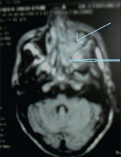
Figure 4: Magnetic resonance imaging brain showing left maxillary (thin blue arrow), ethmoidal and sphenoidal sinusitis (empty arrow)
| Close | |
|
|
|
|
Figure 4: Magnetic resonance imaging brain showing left maxillary (thin blue arrow), ethmoidal and sphenoidal sinusitis (empty arrow)
|
|