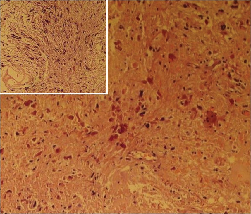
Figure 3: Histopathology showing rosenthal fibers and compact cells with loosely arranged piloid cells (inset)

| Close | |

|
|
|
Figure 3: Histopathology showing rosenthal fibers and compact cells with loosely arranged piloid cells (inset)
|
|