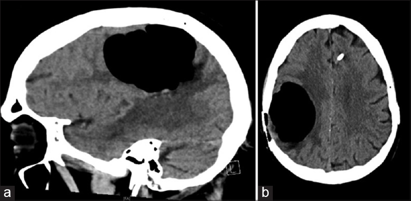
Figure 1: Sagittal (a) and axial (b) sections of computed tomography scan demonstrating pneumocephalus in the index patient

| Close | |

|
|
|
Figure 1: Sagittal (a) and axial (b) sections of computed tomography scan demonstrating pneumocephalus in the index patient
|
|