Copyright restrictions may apply. Please see our Conditions of Use.
| ||||||||||
|
Downloading the PowerPoint slide may take up to 30 seconds. If the slide opens in your browser, select File -> Save As to save it. Copyright restrictions may apply. Please see our Conditions of Use. |
|
|||||||||||

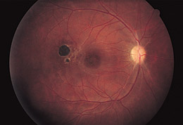
Figure 4. Three months after initial evaluation, there is retinovascular narrowing, central macular pigment change, mild optic nerve pallor, and a new chorioretinal scar in the region of the retinal infiltrate seen in Figure 3. Visual acuity was 20/25 OD.