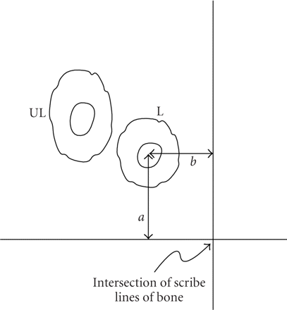
Schematic of method to locate osteons. Multiple orthogonal scribe lines (only one shown) were cut into the polished bone surface.
A labeled osteon was identified under the epifluorescent microscope and the perpendicular distances ( ) from the axes to the center of the Haversian canal were measured. These distances were recorded and once the specimen was
placed under the optics of the indenter microscope, the coordinates to the center of the osteon measured from the intersection
of the scribe lines were entered into the indenter software. This procedure verified the previously identified labeled osteon.
A neighboring unlabeled osteon was identified based on its morphology and proximity to the labeled osteon.


