Effect of tapering internal coronal walls on fracture resistance of anterior teeth treated with cast post and core: In vitro study
- 1Department of Prosthodontics, School of Dentistry, Lebanese University, Beirut, Lebanon
- 2Endodontist, Private Practice, Beirut, Lebanon
- Loubna Shamseddine, Department of Prosthodontics, School of Dentistry, Lebanese University, Mazraa-Daybess Street, Ferdawss Building, First Floor, Beirut 00961, Lebanon. Email: drloubna1{at}hotmail.com
Abstract
When fabricating indirect post and core, internal coronal walls are tapered to remove undercuts and allow a better adaptation. To evaluate the fracture strength of anterior tooth reconstructed with post and core and crowned, with two different taper of internal coronal walls, 6° and 30° to the long axis, two groups of 30 clear plastic analogues simulating endodontically treated maxillary central incisors were prepared. The analogues crowned were subjected to a compressive load with a 1-kN cell at a crosshead speed of 0.05 mm/min at 130° to the long axis until fracture occurred. Data were analyzed by Lillifors and Mann–Whitney tests. Mean failure loads for the groups were as follows: group I 1038.69 N (standard deviation ±243.52 N) and group II 1231.86 N (standard deviation ±368.76 N). Statistical tests showed significant difference between groups (p = 0.0010 < 0.01). Increasing the taper of internal coronal walls appears to enhance the fracture resistance of anterior maxillary teeth post and core reconstructed.
Introduction
Endodontically treated teeth are usually weakened due to loss of dental structure by caries, cavity preparation, root canal instrumentation, and decrease in dentin moisture. They become more susceptible to fractures.1,2 When there is extensive loss of coronal tooth substance, a post is also needed.3 The remaining tooth structure will determine the final restoration planning. In the literature, several factors influence the fracture resistance of endodontically treated teeth: age and sex, position of tooth in the arch, restoration type, periodontal support, and moreover the remaining tooth structure regarding the ferrule size.4⇓⇓–7
Cast post and cores may be used in cases of moderate-to-severe tooth loss structure.8 This treatment modality demonstrated a success rate of 82.6% after 10 years of service.9 Another alternative to post and core reconstruction is the fiber post and composite core. However, this option gives a better prognosis in posterior teeth rather than anterior ones.7,10 Moreover, anterior teeth with cast posts have fracture strength twice the value of fiber posts.11
These findings may be explained by the higher horizontal forces, causing tension stress on anterior teeth in comparison to a more perpendicular compressive force vector for posterior teeth.12,13
It has been established that resistance to fracture is directly related to the amount of remaining tooth structure. While some authors recommended at least 2 mm of ferrule for post placement,14⇓⇓⇓–18 others concluded that a reduction of failures in post endodontic restorations was found when increasing the remaining dentin height, adding to the retention, providing a resistance form, and enhancing the longevity of the restoration.19⇓⇓⇓–23
Moreover, the effect of root canal taper preparation on tooth fracture was also studied.24⇓⇓⇓⇓–29 In an attempt to be conservative, the use of tapered posts requires removing less dentin. Although parallel posts and screw posts are more retentive in the root canal, more dentin removal is required in their post space preparation. This is undesirable as more dentin is removed from the thinner apical and middle aspects of the root canal walls.30
A taper canal preparation of 6°–8° is necessary to ensure an adequate retention while eliminating undesired undercuts.31
The increase in the diameter of posts could increase the stiffness of posts at the expense of the remaining dentin and the fracture resistance of the root.6 In the literature, there is no documentation concerning the effects of taper degree of internal coronal walls on tooth fracture resistance.
The aim of this in vitro study is to evaluate the effect of two different internal coronal taper walls on the fracture resistance of endodontically treated teeth with cast post and core reconstructions. The null hypothesis is that the internal taper of the coronal preparation does not affect the fracture resistance of the teeth.
Materials and methods
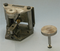
A total of 60 clear acrylic standardized analogues B22X-500 (Kilgore International, Inc., USA) simulating maxillary central incisors were used. These analogues were subsequently mounted on an especially metallic device fabricated for this experiment. This device was made of a base and a mounting block with a hinge axis having the ability to move in a mesiodistal direction. A protractor instrument was related to the base and was used to direct the inclination of the block with precise angulations. The block will precisely hold the analogues in place in a very tight manner. All experimental procedures (see details later) were then realized. At the cementation step, a special cylindrical tube with a central hole was fabricated. This cylinder was held by a plate and mounted on the base parallel and above it. The hole of this cylinder coincided exactly above the hole of the block. It will be used to give access to a metallic rod, in order to cement the post and cores in a uniform manner. This rod with a diameter similar to the fixed cylinder hole would fit and slide along it without friction. This metallic rod was held from one end by a cylindrical plate that would stop the tube once it reached the post and the other end would have a rubber plug that would be used to cement the posts in a homogeneous and reproducible manner (Figure 1)
Preparation of the crown
The crowns were reduced perpendicular to the root axis with an abrasive disc (X928-7 TP; Abrasive Technology, Inc., USA), leaving 5 mm above the cervical area. The axial surface of the tooth was reduced with specific burs (1.5 mm facial ISO no. 806 104 173 544 031, 0.8 mm palatal and proximal ISO no. 806 104 173 544 016) following the cementoenamel junction at the proximal surfaces, to receive a full coverage metal crown.
Endodontic preparation
All analogues were prepared for endodontic treatment with an access cavity of 6° taper, from the incisal edge with 0.8-mm burs (ISO no. 806 104 199 544 016; NTI, Germany). For the ferrule at the proximal side, 2 mm was left. The specimens were divided into two groups of 30 each.
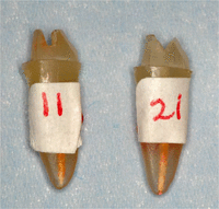
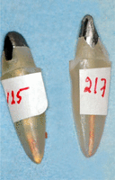
Group I was used as control group, without any modifications, while for group II, facial and palatal residual coronal walls were prepared using a 0.5-mm cylindrical bur and a protractor, deviating the axe of the analogue tooth in a contralateral direction to the internal wall that needs to be prepared in order to create a 30° internal taper to the long axis of the tooth (Figures 2 and 3).
The preparation of root canal was executed with the ProTaper system until 18 mm was reached (conventional step-back technique). ProTaper is a progressive tapered instrument conceived for shaping and finishing root canals. Two files (S1 and S2) were used for shaping and two files (F1 and F2) were used for finishing (Dentsply Maillefer Instruments, Switzerland). Gutta-percha was laterally condensed with a manual spreader (Kerr, W 0697840 and W 0693510).
Post and core fabrication
The post spaces were prepared for each canal with no. 3 Gates-Glidden drill (Dentsply Maillefer A0008 240 005 00) and a 1.1-mm-diameter Largo (Dentsply Maillefer A0008 230 002 00 to A0008 230 003 00) to within 7 mm of the apex to ensure the apical seal.
For each tooth, a 1.25-mm plastic burnout pattern (Coltène Whaledent) was inserted into canal space, and a custom post and core was constructed using post and core wax pattern (Harvard Blue-Wax; Richter & Hoffman, Harvard Dental GmbH, Germany).
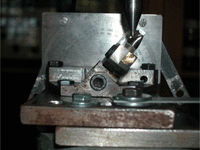
The core dimensions were kept the same for the two groups using silicone index. The post and cores were cast in a Nickel Chrome alloy. Cementation was realized using spiral paste filler (Dentsply Maillefer Instruments), under a static load of 1.5 kg for 15 min, with zinc phosphate cement (SpofaDental Adhesor®) mixed according to manufacturer’s directions (Figure 4).
Cast crown fabrication
After removing cement excess, an overcastting crown was waxed and adapted directly to the analogue using the silicone index done after predesigned indentation on the lingual aspect of the analogue. The wax pattern was then invested and cast using the Nickel Chrome metal and then cemented with zinc phosphate under the same static load that was applied for the post and core cementation.
Fracture testing
For the two groups, fracture strength test was done at the laboratory of the Mechanical Engineering Department at the American University of Beirut (Lebanon). The testing device was a tension and compression system YL Universal Testing Machine (YLUTM). The device was fully controlled by computer. Certified and calibrated by the National Institute of Sciences and Techniques (NIST) known as National Bureau of Standards, the testing machine allowed an error of 0.04% for maximal load 10,000 kg, 0.01% for repetitive maximal load 10,000 kg, resolution of displacement 0.01 mm (10 µm), and accurate speed of 0.01% of full scale.
The analogues crowned were subjected to a compressive load with a 1-kN cell at a crosshead speed of 0.05 mm/min at 130° to the long axis until fracture occurred (Figure 5). To analyze results, statistical software (SPSS 17; SPSS Inc., USA) was used. Averages and standard deviations (SDs) were established. Statistical tests of normality (Lillifors) were used to check whether data match a particular distribution such as the normal distribution or the exponential distribution.
Nonparametric Mann–Whitney test was used to test the null hypothesis for the strength between groups. Probability value less than p < 0.05 was set. For the displacement values, the nonparametric Kruskal–Wallis test was used to compare independent groups (p ≤ 0.05).
Results
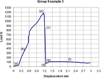
All specimens showed a similar curve with seven values as presented in Figure 6.
Curve represented for each specimen under compression: (a) artifact when the compressive force is absorbed for the final seating of the stem into the palatal indentation, (b) increased compressive force to the point of breakage of the root or initial failure, (p) initial failure or fracture root, (c) decompression after initial failure, (d) detachment pieces, (e) slight increasing compression due to friction of post on root walls, and (f) horizontal line represents a full decompression when post is completely removed.
Note that value “(a)” is an artifact corresponding to the final compression force when the rod hits the indentation. This value was subtracted from the total values “(a)” to “(p).”
Statistical results are summarized in Table 1. Greater resistance was observed in the group with 30° to the long axis direction of internal coronal walls (1231.86 ± 368.76 N) than in the group with 6° taper (1038.69 ± 243.52 N). The Mann–Whitney strength test revealed significantly greater resistance in the group with 30° preparation taper (p = 0.0010) than in the group with 6°. The Kruskal–Wallis test revealed that there is no significant difference for the displacement between the two groups.
Summary of the statistical results.
Discussion
To standardize all parameters affecting the fracture behavior of teeth such as section and dimension of posts,32⇓⇓⇓⇓–37 clear acrylic analogues were used in this study. These results have a limited clinical value as the load fracture found does not reflect the mouth situation (because of the differences between natural and acrylic teeth in adhesion behavior38,39 and fracture strengths40). The major objective of this study is to find which preparation design has a better resistance force to fracture, while a specific value can only express the mechanical properties of plastic analogues.
As for the device, Milot and Stein used almost the same device and teeth. Their study was about the effect on fracture resistance when comparing margin preparation design in relation to several metallic post and core systems. Our device had the advantage of being able to have angulated positions around an axis and our study had a more specific goal: the internal axial wall design preparation.41
The taper of the internal wall was 6°. It corresponds to the taper of the bur used. The 30° tapering was chosen to preserve the maximum of remaining tooth structure while trying to have a good seating of the posts. This decision was hypothetical and based on a study done for crown preparation design.42 No study was found in regard to internal wall design.
There was a significant difference (p = 0.001) between internal wall tapering preparation with a 30° taper, when compared to the 6° taper. It was shown that the 30° taper gave a greater resistance to fracture.
These results would be due to a combination of a better surface contact post/analogue and a more favorable stress distribution. It is generally accepted that cast post and core success is related to an accurate reproduction of the canal shape and to a very well seated core in order to distribute forces uniformly and maximize the mechanical behavior of the final restoration.43⇓–45
When a post is supported with core and coronal coverage, stress distribution becomes a larger function of the post geometry and “fit.”6,34,46,47 This is in line with our findings that the 30° internal wall preparation gave better mechanical results. Wall tapering increase was also found to improve the wax pattern filling of cast post and core, thus increasing the fit of the cast post and core.
To ensure a perfect seating of cast post and core, some authors recommended the creation of cement escape like venting mechanism to be incorporated in wax pattern, before or during cementation; in this case, a bur should be used to create this venting mechanism.31,48
Group II results in this study could be explained by a uniform stress distribution between post and the canal as the cement layer provides a buffer zone.49⇓–51 It has been shown in the crown cementation procedure that wall tapering affects zinc phosphate filtration phenomenon and escapement at cementation procedure52,53 and alleviates hydrostatic pressure during cementation.8,53⇓–55 The taper degree was also shown to improve the cementation and seating of cast post and core similar to the cementation of crowns.
In group II, 30° of wall tapering leads to a geometrically bigger contact surface and consequently a bigger effective surface. Stresses are defined as force per unit area on the surfaces of a differential cube. These stresses are described as tensions or compressions. For other coordinate frames, such as cylindrical or spherical coordinates, the shape of the test volume contains curved surfaces, but the nature of the stresses is similar. Mathematically, normal stress is most commonly designated with Greek letter sigma (σ). Stress is inversely proportional to the surface contact: σ = F/S. Since the surface in group II increases, stress in group II decreases.
The decrease in fracture resistance of group I is in agreement with the study by Giovani et al. in 2009, which stated that as the mass volume of posts decreases, the absorption of forces by post system also decreases to a considerable degree. The result being that the mass volume will transfer the forces more efficiently to a less rigid tooth dentin, thus endangering the tooth structure with an increase in root catastrophic fracture.33
Conclusion
Within the limitations of this study, for cast post and core reconstruction, the following can be concluded:
-
The increase in the taper of the internal residual coronal walls enhances the fracture resistance of anterior teeth.
-
In situations where preparation has more than 2 mm of ferrule, the thickness of the remaining coronal substances does not affect the fracture strength.
Additional in vivo studies should be performed to confirm these findings before the clinical use of this preparation is recommended.
Acknowledgments
The authors would like to thank Jose Johann Chidiac (Postgraduate studies in Prosthodontics, School of Dentistry, Lebanese University) for his help in editing the manuscript.
Article Notes
-
Declaration of conflicting interests The authors declare that there is no conflict of interest. As in vitro study either the ethical committee or the patient consent is not applicable.
-
Funding This research received no specific grant from any funding agency in the public, commercial, or not-for-profit sectors.
- Received March 2, 2014.
- Accepted July 23, 2014.
- © The Author(s) 2014
This article is distributed under the terms of the Creative Commons Attribution-NonCommercial 3.0 License (http://www.creativecommons.org/licenses/by-nc/3.0/) which permits non-commercial use, reproduction and distribution of the work without further permission provided the original work is attributed as specified on the SAGE and Open Access page (http://www.uk.sagepub.com/aboutus/openaccess.htm).

