- Institution: Stanford Univ Med Ctr Lane Med Lib/Periodical Dept/Rm L109
- Sign In as Member / Individual
Signaling with Phosphoinositides: Better than Binary

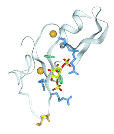
Figure 4.
Structure of the PI3P-bound FYVE domain of EEA1. The ribbon indicates the four β-strands and the C-terminal helix; the two zinc ions are shown as orange spheres. The side chains of an exposed threonine and valine that insert into the membrane are shown in orange at the bottom. The pairs of arginines (blue) that recognize the 3- and 1-phosphates of the inositol ring (yellow and red) are drawn above and below the two conserved histidines (green), respectively. (See (32).)


