- Institution: Stanford Univ Med Ctr Lane Med Lib/Periodical Dept/Rm L109
- Sign In as Member / Individual
Expression Array Technology in the Diagnosis and Treatment of Breast Cancer

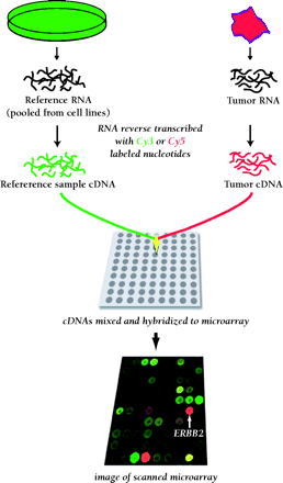
Schematic of microarray technique. RNA from a tumor sample and reference RNA (made commercially from pooled cell cultures to represent the majority of known genes) are reverse transcribed and labeled with different fluorescent dyes. The mixture is hybridized overnight to a microarray. The hybridized microarray is then scanned at two wavelengths and the intensities of red and green fluorescence are measured at each spot on the microarray. The red-to-green ratio reveals the abundance of RNA expressed by the tumor sample relative to the reference sample for every one of the 42,000 cDNA clones on the array. This technique provides a comparative measure of the global gene expression of the tumor sample.


