- Institution: Stanford Univ Med Ctr Lane Med Lib/Periodical Dept/Rm L109
- Sign In as Member / Individual
Neuropeptide S: A New Player in the Modulation of Arousal and Anxiety

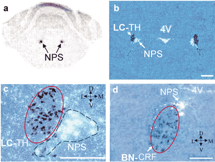
Localization of NPS precursor mRNA expression in the rat brainstem by in situ hybridization. A. Autoradiogram showing strong labeling of NPS-expressing cells in the LC area of the rat brainstem. B. Double in situ hybridization for NPS precursor (white) and tyrosine hydroxylase (TH, dark blue) demonstrates that the NPS-producing cells are not colocalized with noradrenergic LC neurons (LC-TH), but rather form a separate cluster in close vicinity. (4V, fourth ventricle.) C. Higher magnification of (B). The cluster of noradrenergic LC neurons (LC-TH) is circled with a solid red line; the NPS cluster is marked with a broken green line. D. Double in situ hybridization for NPS precursor (white) and corticotropin-releasing factor (CRF, dark blue). CRF-positive cells form the ovoid-shaped Barrington’s nucleus (BN-CRF; solid red line). Scale bar in B is 500 μm, and 250 μm in C and D. Crossed arrows denote orientation of slides: D, dorsal; V, ventral; L, lateral; M, medial. Photomicrographs are adapted from (23).


