|
Show all abstracts Show selected abstracts Add to my list |
|
| REVIEW ARTICLES |
|
|
|
Autotransplantation of cryopreserved teeth: A review |
p. 1 |
Siddharth Sonwane, B Sunil Kumar, P Ganesh, Vishwanath Patil, RGK Shett
DOI:10.4103/2321-3825.147972 Autotransplantation of teeth, if carried out successfully, ensures that alveolar bone volume is maintained due to physiological stimulation of the periodontal ligament. Autotransplantation has been carried out for many years, but with varying success rates. As a result, it is seldom regarded as an appropriate treatment option for patients. Autotransplantations of teeth are widely used in cases of severe impactions, early loss of permanent teeth, or congenital aplasia. However, sometimes, patients may not have a donor tooth available due to previous extraction. To solve such problems, teeth cryopreservation systems have been developed. There are many clinical reports and animal experiments showing the efficacy of teeth cryopreservation. Hence, unnecessary wisdom teeth, supernumerary teeth, and healthy premolars extracted by orthodontic treatment should be used as donor teeth for replacing a missing tooth in the future. In this review, the biological properties of cryopreserved teeth, clinical application of missing teeth are discussed. |
| [ABSTRACT] [HTML Full text] [PDF] [Mobile Full text] [EPub] [Sword Plugin for Repository]Beta |
|
|
|
|
|
|
| ORIGINAL ARTICLES |
 |
|
|
|
Study of the effects of oral irrigation and automatic tooth brush use in orthodontic patients with fixed appliances |
p. 4 |
Dolly Patel, Falguni Mehta, Ipsit Trivedi, Nishit Mehta, Unnati Shah, Vijay Vaghela
DOI:10.4103/2321-3825.147973 Objective: To compare effectiveness of: 1) Conventional tooth brush alone (control) 2) Powered tooth brush alone 3) Conventional tooth brushing with oral irrigation device 4) Powered tooth brush with oral irrigation device, as home use oral hygiene methods in adult fixed orthodontic patients. Materials and methods: Sixty orthodontic patients with fixed orthodontic appliances were divided into four study groups: (A) brushing with automatic tooth brush twice daily (n = 15); (B) oral irrigation with manual toothbrushing, (n = 15); (C) oral irrigation with automatic tooth brushing, (n = 15); (D) control group with continued normal tooth brushing only, (n = 15). Gingival and plaque indices, bleeding after probing, and gingival sulcus depths were assessed at baseline, 1-month, and 2-month periods. Results: Paired t test was used for within group analysis. Tukey's honestly significant difference (HSD) statistical analysis was used for the inter-group multiple comparisons. Level of significance was at P < 0.05. Within group comparison reveal that there are no statistically significant differences between the groups regarding reductions in gingival index mean scores in all 3 time durations except for group C (P = 0.68) and group D (P = 0.93) in the time period of 1 month to 2 months. After 1 to 2 months use of the automatic tooth brush, there was a significant reduction in plaque when compared with the control group who used only the manual tooth brush (P = 0.04). For this population of orthodontic patients, powered brushes alone or along with oral irrigation do not have additional beneficial effect when comparisons are made with other groups. Conclusion: All plaque control methods evaluated in the study provides significant improvement in reduction of plaque accumulation and gingival inflammation. Powered brushes alone or along with oral irrigation do not seem to be additionally beneficial when comparisons are made with other groups. |
| [ABSTRACT] [HTML Full text] [PDF] [Mobile Full text] [EPub] [Sword Plugin for Repository]Beta |
|
|
|
|
|
  |
Pharyngeal airway parameters in subjects with Class I malocclusion with different growth patterns |
p. 11 |
Dipti Shastri, Pradeep Tandon, Amit Nagar, Alka Singh
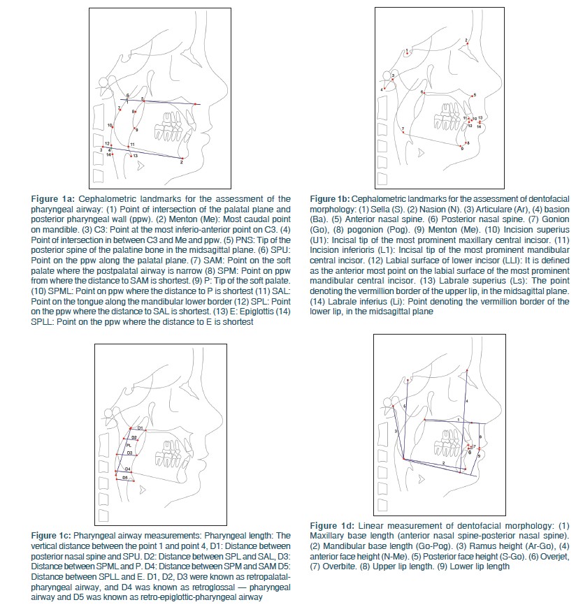
DOI:10.4103/2321-3825.146359 Objectives: (1) To test the null hypothesis that there are no significant difference in the pharyngeal airway in subjects with Class I malocclusion with different growth patterns. (2) To test the null hypothesis that there are no significant difference in dentofacial structure in subjects with Class I malocclusion with different growth patterns. Materials and Methods: Lateral cephalometric radiographs of 120 skeletally Class I were separated into three groups according to the SN-MP angle. Lateral cephalometric radiographs of 39 low angle, 45 high angle and 36 normal angle were examined. Group difference were analyzed with analysis of variance (ANOVA) and the Tukey test, at the P < 0.05 level. Results: For pharyngeal airway measurements statistically significant difference were found in pharyngeal airway length, and D5 (retroepiglottal) pharyngeal width. No statistically significant sagittal pharyngeal (D1-D5) parameters difference were determined between low angle and normal angle subjects. High angle subjects had lower sagittal pharyngeal D2 (retropalatal) and D5 (retroepiglottal) parameters than those with low and normal angle, additionally in high angle subjects had lower D1 (retropalatal) and D4 (retroglossal) parameters than those with normal angle subjects. According to ANOVA only 1 out of 9 dentofacial measurements showed not statistically significant difference among different growth patterns. Conclusion: The null hypothesis was rejected. Significant difference in pharyngeal airway measurements and dentofacial morphology of Class I subjects with different growth patterns were identified. |
| [ABSTRACT] [HTML Full text] [PDF] [Mobile Full text] [EPub] [Sword Plugin for Repository]Beta |
|
|
|
|
|
  |
Jasper Jumper and activator-headgear combination: A comparative cephalometric study |
p. 17 |
Emine Kaygisiz, Tuba Tortop, Sema Yuksel, Selin Kale Varlik, Erdal Bozkaya
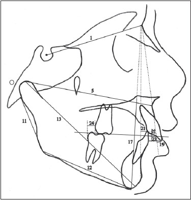
DOI:10.4103/2321-3825.146360 Aim: The aim of this study was to evaluate and compare the skeletal and dentoalveolar effects of Jasper Jumper (JJ) and activator-headgear (AcHg) combinations and an untreated control group. Materials and Methods: The sample comprised 37 Class II high-angle patients. Twenty of them (mean age: 12.4 ± 0.61 years) were treated with JJ and 17 of them (mean age: 10.9 ± 0.74 years) were treated with AcHg. Mean treatment time was 5 months for the JJ group and 11 months for the AcHg group. Control group consisted of 20 Class II high angle patients (mean age: 10.4 ± 0.41 years) and mean observation period was 10 months. Results: Co-A showed significant increase in the JJ and control groups while SNA angle decreased significantly in only JJ group. Increase in SNB angle in AcHg group was significantly greater than in the JJ and control groups. In the JJ group, mandibular incisors protruded significantly. Conclusion: Both the AcHg and JJ treatments had restraining effect on maxillary growth, but stimulated significant mandibular growth. Anteroposterior discrepancy was corrected in the AcHg group mostly by the mandibular growth compared to JJ treatment. Maxillary incisors were retroclined in the AcHg group while mandibular incisors were proclined in the JJ group. |
| [ABSTRACT] [HTML Full text] [PDF] [Mobile Full text] [EPub] [Sword Plugin for Repository]Beta |
|
|
|
|
|
  |
Evaluate the influence of panel composition on facial attractiveness |
p. 25 |
Rajkumar Maurya, Ankur Gupta, Jaishree Garg, Chandresh Shukla
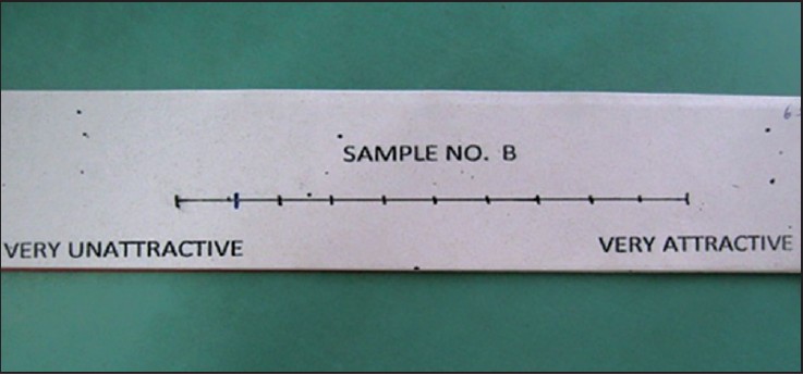
DOI:10.4103/2321-3825.146361 Introduction: The study was conducted to evaluate the influence of professional background, age and gender, of panel members on their evaluation of the facial attractiveness of adolescents. Materials and Methods: A panel of 20 adult laymen 20 professionals (orthodontist and maxillofacial surgeons) evaluated photographic sets (one frontal and one lateral view) of 170 adolescents (76 boys and 94 girls) on a visual analogue scale (VAS) in relation to a reference set of photographs. The effects of the characteristics of the panel members on the VAS scores for boys and girls separately, as well as their interactions, were evaluated by multilevel models. Statistical Analysis: The influence of professional background, age and gender on the VAS scores for the boys and the girls separately and their possible interactions were tested within the framework of multilevel models. Student's t-test for equality of means and Levene's test for equality of variances were performed. Conclusion: The multilevel model and first-order interactions revealed that laymen rated adolescents as more attractive than professionals. Male laymen rated the boys and girls significant more attractive than male professionals. Young laymen rated boys and girls significantly more attractive than young professionals. Young laymen rated girls significantly more attractive than old laymen. |
| [ABSTRACT] [HTML Full text] [PDF] [Mobile Full text] [EPub] [Sword Plugin for Repository]Beta |
|
|
|
|
|
  |
Two-dimensional to three-dimensional: A new three-dimensional cone-beam computed tomography cephalometric analysis |
p. 30 |
Raghu Devanna
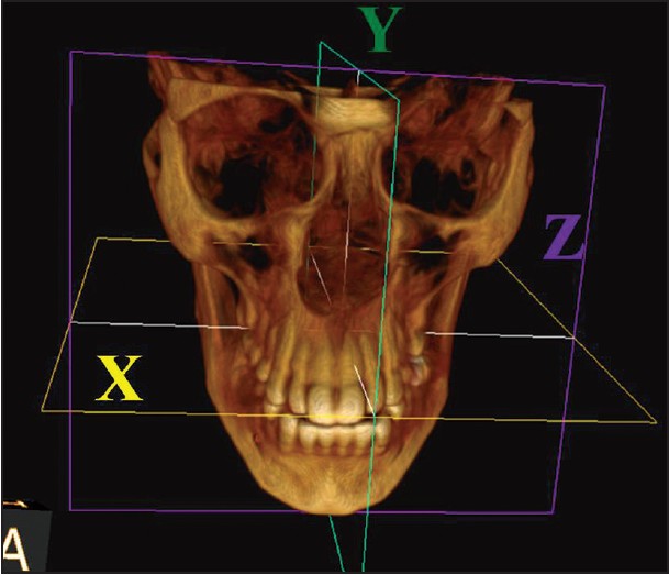
DOI:10.4103/2321-3825.146356 Objectives of the Study: were (1) to develop a three-dimensional cephalometric analysis scheme applicable to assessing dentofacial deformities; and (2) to create a normative database of three-dimensional cephalometric measurements for adult North Karnataka population. Materials and Methods: A cross-sectional study was conducted on 40 male and 40 female adults with normal balanced facial profile and occlusion. Cone-beam computed tomography (CBCT) images obtained in digital imaging and communications in medicine format and the anatomic Cartesian three-dimensional cephalometric reference system according to Swennen et al. was used to standardize the reference planes. Cephalometric analysis was performed using various landmarks. New three-dimensional cephalometric analyses appropriate for orthognathic surgery as well as new parameters were used in this study. Results: The cephalometric norms generated in this study were comparable with those reported in the literature for conventional two-dimensional cephalometric analysis and unique features of North Karnataka population. The results showed significant differences between males and females in most of the facial measurements (P < 0.0001). Conclusion: This is the first database of three-dimensional cephalometric norms based on CBCT of the North Karnataka population. Norms generated were comparable with those reported in the literature with the conventional two-dimensional cephalometry: More accurate and reliable. Moreover, three-dimensional cephalometric analysis has the potential of incorporating new measurement methods that are difficult if not impossible in two-dimensional cepholmetric analysis. This method of cephalometric analyses can be useful in diagnosis and treatment planning for patients with dentofacial deformities. |
| [ABSTRACT] [HTML Full text] [PDF] [Mobile Full text] [EPub] [Citations (3) ] [Sword Plugin for Repository]Beta |
|
|
|
|
|
  |
Evaluation of self-etching primer and erbium-doped yttrium aluminum garnet laser on bracket bond strength: An in-vitro study |
p. 38 |
Pawankumar Dnyandeo Tekale, Ketan K Vakil, Jeegar K Vakil
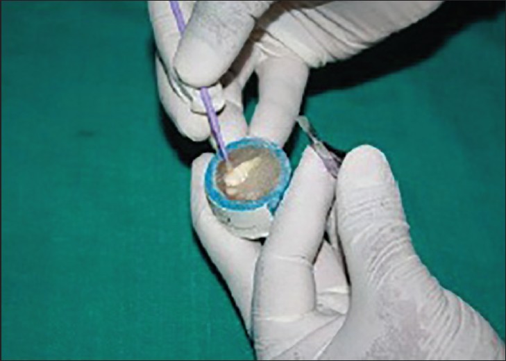
DOI:10.4103/2321-3825.146355 Introduction: The purpose of this study was to evaluate and compare the shear bond strength, the adhesive remnant scores and surface characteristics of the teeth prepared for bonding with erbium-doped yttrium aluminum garnet (Er:YAG) hard tissue laser, self-etching primer (SEP) and phosphoric acid etching. Materials and Methods: Seventy-eight human premolars, extracted for orthodontic purposes were randomly divided into three groups, enamel was irradiated with 37% phosphoric acid in Group-1, with SEP in Group-2 and with Er:YAG laser at 1.5-W in Group-3. After surface preparation standard edgewise stainless steel premolar brackets were bonded; one tooth in each group was not bonded and was examined under a scanning electron microscopic. The brackets were debonded 24 h later; shear bond strengths were measured, and adhesive remnant index scores were recorded. Results: Statistically significant differences were found between phosphoric acid etching, SEP, 1.5-W laser irradiation. Adhesive remnant scores were compared with the Chi-square test, and statistically significant differences were found between all groups. Conclusions: The mean shear bond strength obtained with 37% phosphoric acid etching, SEP and Er-YAG laser etching were clinically acceptable. SEP produces a more conservative etch pattern and more shear bond strength than phosphoric acid. More adhesive was left on the SEP treated enamel as compared to enamel treated with acid and laser etching. |
| [ABSTRACT] [HTML Full text] [PDF] [Mobile Full text] [EPub] [Sword Plugin for Repository]Beta |
|
|
|
|
|
  |
Assessing reliability of mandibular planes in determining gonial angle on lateral cephalogram and panoramic radiograph |
p. 45 |
Thilagarani , Purnima V Nadkerny, Danyasi Ashok Kumar, Vidyesh D Nadkerny
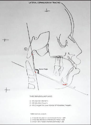
DOI:10.4103/2321-3825.146358 Introduction: Gonial angle is used widely in orthodontic tracing and is a significant indicator to diagnose the growth pattern in patients. Lateral cephalograms and panoramic radiographs both effectively determine the gonial angle. A definite variation exists when the gonial angle is determined using three different mandibular planes as described by Tweed, Steiner, and Down, respectively. The aim of this study was to evaluate, which gonial angle (obtained from Tweed's, Steiner's or Down's mandibular plane) on lateral cephalogram has the value closest to that obtained on a panoramic radiograph. Materials and Methods: A total of 300 patients between 12 and 29 years of age were selected. Panoramic and lateral cephalometric radiographs were obtained. On panoramic radiograph, gonial angle was determined by tangent of inferior border of mandible and most distal aspect of ascending ramus and condyle and compared with the value of each gonial angle obtained using three mandibular planes as described by Tweed, Steiner and Downs on lateral cephalogram. Levene's and Independent samples t-test were done for comparison. Results: Difference between gonial angles determined by Tweeds mandibular plane on lateral cephalogram and panoramic radiograph was statistically insignificant (P > 0.05). Conclusion: Of the three commonly used mandibular planes in measuring gonial angles, the value obtained with Tweeds mandibular plane seems to be more reliable. |
| [ABSTRACT] [HTML Full text] [PDF] [Mobile Full text] [EPub] [Sword Plugin for Repository]Beta |
|
|
|
|
|
  |
Photographical evaluation of smile esthetics after extraction orthodontic treatment |
p. 49 |
Veerendra Prasad, Pradeep Tandon, Vijay Prakash Sharma, Gulshan Kumar Singh, Rana Pratap Maurya, Vinay Chugh
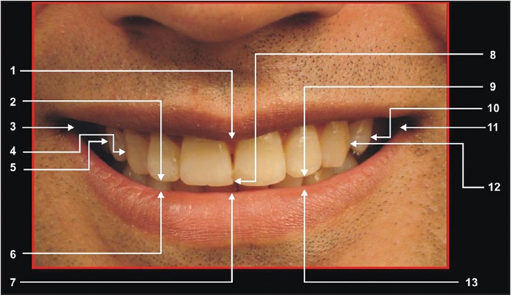
DOI:10.4103/2321-3825.147976 Aims: To evaluate and compare the smile esthetics in orthodontically treated subjects and subjects with an esthetically pleasing smile. Materials and Methods: Frontal smiling photographs of 80 subjects in the age group of 18-25 years (mean age of 21.97 years) were taken and divided into Group I (having an esthetically pleasing profile and normal occlusion) and Group II (orthodontically treated). Each Group had 40 subjects, who were further divided into male and female subgroups. Eight transverse and three vertical linear measurements were taken on the frontal photographs and eight ratios were derived. Esthetic scores and other variables were also obtained. The data so obtained were subjected to statistical analysis. Results: All seven ratios did not show any statistically significant differences in both the groups except for ratio 5 (<0.05) in Group IIb. No statistically significant differences were found in the variables of the upper lip curvature, visible marginal gingiva or visible mandibular teeth, except in the visible maxillary first molar (<0.05) for males. The esthetic score showed statistically higher values for males (<0.05) and females (<0.001) in Group I. Lay persons rated significantly higher mean values for esthetic scores in Group Ia (<0.05), Group Ib (<0.001), and Group IIb (<0.01). There were no significant correlations found between the esthetic scores and the seven ratios for both the groups. Conclusion: (1) Females had a more interpremolar/smile width ratio. (2) A greater positive upper lip curvature was found in Group I males and females and was rated higher for esthetic score. (3) The visible maxillary first molar was more in Group II males and females and rated lower for esthetic score. (4) Esthetic scores rated by lay persons were higher for all the subjects. |
| [ABSTRACT] [HTML Full text] [PDF] [Mobile Full text] [EPub] [Sword Plugin for Repository]Beta |
|
|
|
|
|
|
| CASE REPORTS |
 |
|
|
  |
Orthodontic camouflage treatment in skeletal Class II patient |
p. 57 |
MB Raghuraj, Rajat Scindhia, Vivek Amin, Sandeep Shetty, Rohan Mascarenhas, Nandish Shetty
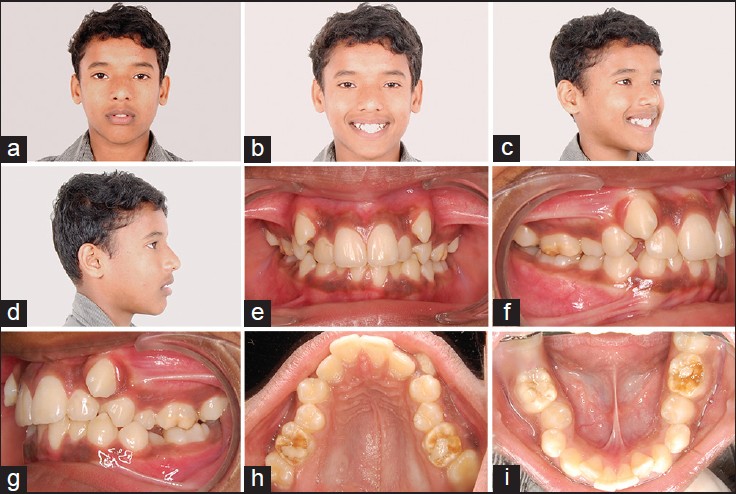
DOI:10.4103/2321-3825.146354 Orthodontic camouflage is a method of correcting malocclusion without involving the correction of skeletal problem. Planned extraction of some teeth will help us achieve favorable dental occlusion. The challenge lies in proper diagnosis and case selection so as to decide on dental camouflage as a treatment option in skeletal discrepancy cases. Case of Class II malocclusion with severe crowding, vertical growth pattern and Class II skeletal base with ANB 6° has been discussed. Treated with four premolar extractions and finished the case with Class I canine and molar relationship. Planned extraction of indicated teeth to bring about dental compensation and camouflage the underlying skeletal discrepancy gives an overall improvement in facial esthetics, occlusion and also satisfaction to the patient. |
| [ABSTRACT] [HTML Full text] [PDF] [Mobile Full text] [EPub] [Sword Plugin for Repository]Beta |
|
|
|
|
|
  |
Management of maxillary lateral incisor: Canine transposition along with maxillary canine impaction on the contralateral side |
p. 61 |
Veerendra Prasad, Pradeep Tandon, Gyan Prakash Singh, Rana Pratap Maurya
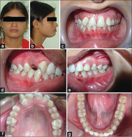
DOI:10.4103/2321-3825.146357 This is a case of 24 years 5-month-old female whose chief complaint was irregularly arranged front teeth. The clinical and radiographical examination revealed pleasing profile, Angle's Class I molar relationship, complete transposition of maxillary right lateral incisor and canine along with impacted left maxillary canine and retained deciduous canine. Maximal band tension 0.022″ × 0.028″ appliance was placed. Transposed right maxillary canine and lateral incisor as well as impacted left maxillary canine was aligned in the dental arch. The total treatment time taken was 24 months, which was followed by a lingual-bonded canine-to-canine retainer. |
| [ABSTRACT] [HTML Full text] [PDF] [Mobile Full text] [EPub] [Sword Plugin for Repository]Beta |
|
|
|
|
|
  |
Bone-anchored maxillary protraction followed by fixed appliances in skeletal Class III malocclusion: A report of two cases |
p. 65 |
Emine Kaygisiz, Erdal Bozkaya, Sema Yuksel, Erkan Erkmen, Mustafa Sancar Atac
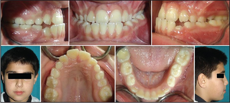
DOI:10.4103/2321-3825.146362 Maxillary protraction is recommended for patients with skeletal Class III malocclusion diagnosed with maxillary deficiency. The major goal of the protraction is to correct maxillary discrepancy with a skeletal alteration rather than dental movements. However, loss of the dental anchorage has been reported beside the skeletal effects with facemask therapy using tooth-borne anchorage. The use of miniscrews and miniplates became popular as an anchorage instead of conventional tooth-borne appliances. Studies evaluating the second-phase treatment results and stability of bone-anchored maxillary protraction are needed in literature. This case report presents the treatment of 11 and 12-year-old two boys with Class III malocclusion due to maxillary deficiency. Both cases were treated with fixed appliances subsequent to maxillary protraction with miniplates. Maxillary skeletal effects were increased, and undesired dentoalveolar effects were reduced by the facemask therapy with skeletal anchorage. The occlusion and the facial profile were effectively improved, with good stability after fixed appliances. |
| [ABSTRACT] [HTML Full text] [PDF] [Mobile Full text] [EPub] [Sword Plugin for Repository]Beta |
|
|
|
|
|
  |
Treatment of Class III malocclusion in a young adult with reverse pull face mask  |
p. 70 |
Zeeshan Iqbal Bhat, Jayesh S Rahalkar, Sonali Deshamukh, Charu Dutta Naik
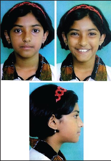
DOI:10.4103/2321-3825.147986 Class III malocclusions are usually growth-related discrepancies, which often become more severe until growth is complete. This case report describes the treatment of a young girl aged 11 years 3 months who had a skeletal Class III malocclusion with a flattening of mid facial region. She also had constricted maxillary arch and high labially placed canine. The treatment plan included a slow palatal expansion, reverse pull facemask appliance, and fixed edgewise appliances. The treatment resulted in skeletal Class I and dental Class I molar and canine occlusion, an ideal overjet, overbite, incisor angulation and facial esthetics was greatly improved after 21 months of treatment. Stability of the treatment result was excellent in 1 year 5 months follow-up at the age of 14 years and 7 months. |
| [ABSTRACT] [HTML Full text] [PDF] [Mobile Full text] [EPub] [Sword Plugin for Repository]Beta |
|
|
|
|
|
  |
Non-surgical management of unilateral cleft lip and palate in growing patient |
p. 76 |
Falguni Mehta, Renuka Patel, Vaibhav Gandhi, Manop Agrawal
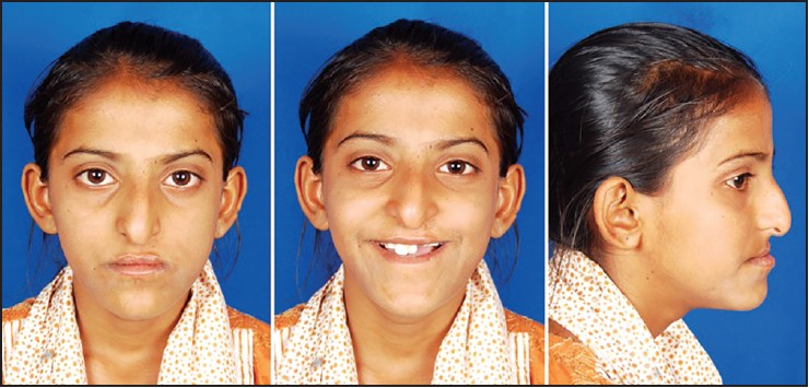
DOI:10.4103/2321-3825.147987 Cleft lip and palate (CLCP) is one of the most common congenital deformities of the craniofacial complex. It has multifactorial etiology with skeletal, dental, soft tissue defects; functional defects such as speech, swallowing, hearing; in addition to psychological and social implications along with facial disfigurement. Thus, the treatment approach is also multidisciplinary involving orthodontist, plastic surgeons, ear-nose and throat surgeon, oral and maxillofacial surgeon, prosthodontist, speech therapist, etc. This case report describes a nonsurgical approach for treating the CLCP patient with rapid maxillary expansion followed by Facemask therapy for protraction of maxilla during the mixed dentition phase to correct the developing malocclusion. |
| [ABSTRACT] [HTML Full text] [PDF] [Mobile Full text] [EPub] [Sword Plugin for Repository]Beta |
|
|
|
|
|